

This post will consist of 15 gross pathology pictures. We will also provide descriptions of each disease. The descriptions are concise and simple to comprehend. For some of the medical terms, we added links to reputable sites that provide more detailed information. Before you proceed, we must warn you that some of the images are not recommended for people with a weak stomach.
What Does The Term “Gross Pathology” Mean?
Wikipedia defines gross pathology as macroscopic (objects with a large enough size to be visible virtually with the naked eye) manifestations of disease that can be seen in organs, tissues and body cavities. Anatomical pathologists will often use this particular term to refer to diagnostically useful findings that they had made during the gross examination (a process in which pathology specimens are first inspected with the naked eye for diagnostic information purposes, and then later processed for a microscopic examination) segment of surgical specimen processing or an autopsy.
15 Up-Close & Detailed Pictures
Peritoneal Cyst
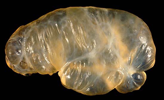 This is a benign 8-centimeter cyst found floating in the peritoneal cavity following a hysterectomy.
Fibrous Plaque On The Thoracic Diaphragm
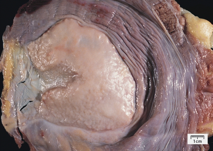
This is a fibrous plaque on the thoracic diaphragm. It was found in an asbestos worker.
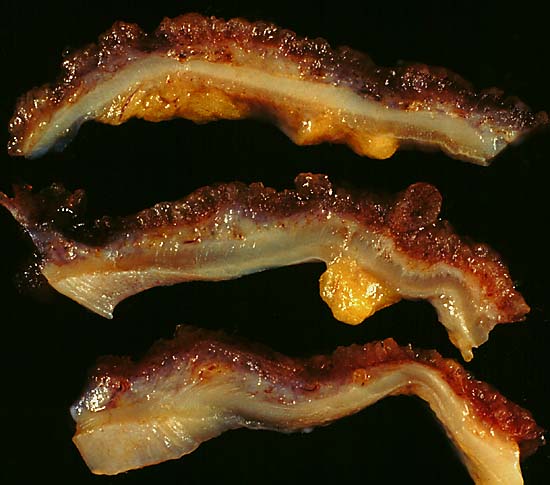
Chronic Ulcerative Colitis

Ulcerative Colitis is an inflammatory bowel disease. It affects the lining of the colon and rectum.
Close-Up View Of Chronic Ulcerative Colitis
Ameloblastoma
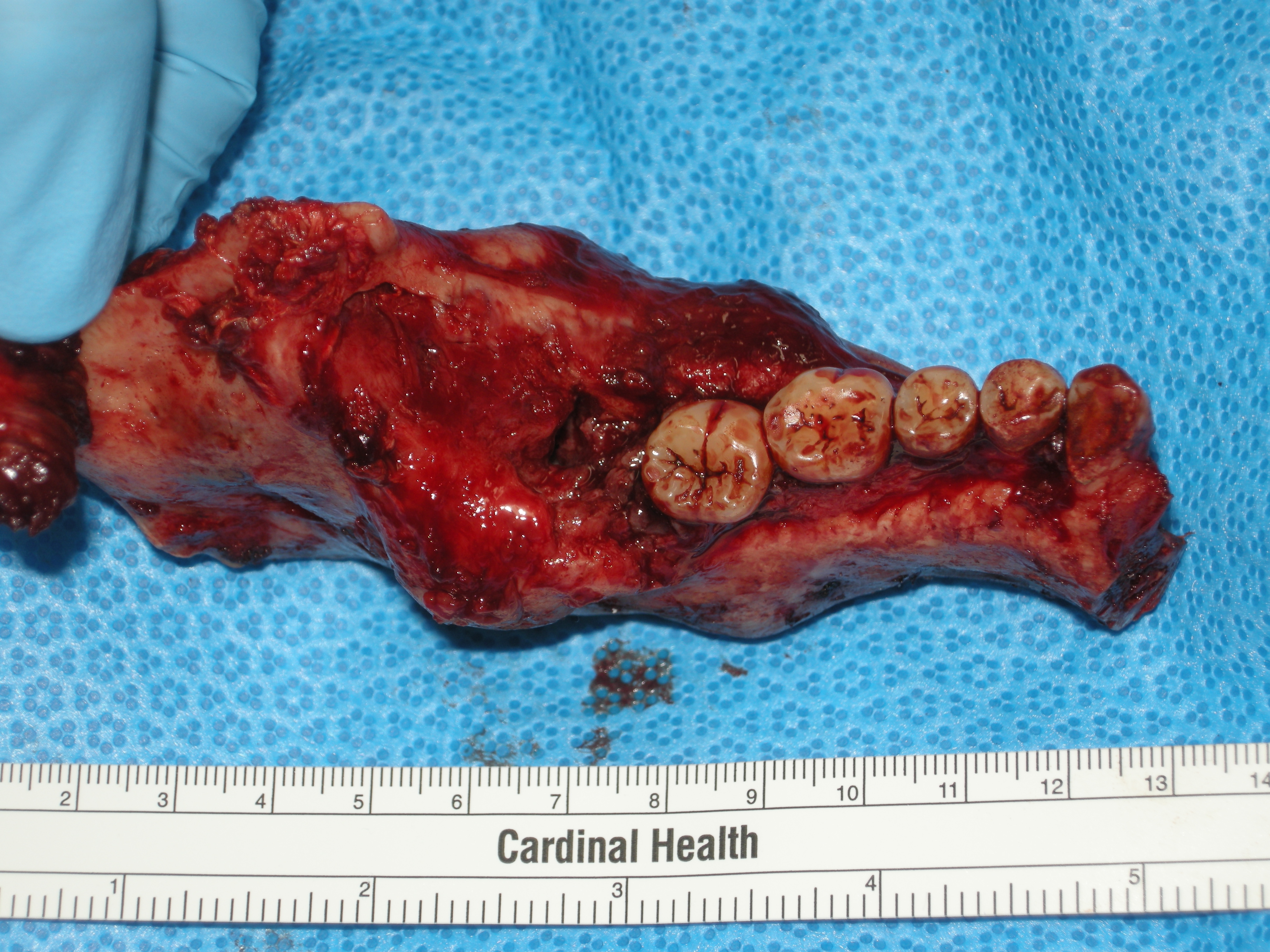
The above picture shows the resected (cut out) left half of a patient’s mandible containing an ameloblastoma. An ameloblastoma is defined as a rare and benign tumor of odontogenic (teeth formation) epithelium. The tumor commonly appears in the lower jaw more than the upper jaw.
Man With Ameloblastoma Of The Mandible

“Ameloblastoma” by Camazine is licensed under CC BY 3.0
Child With A Massive Ameloblastoma
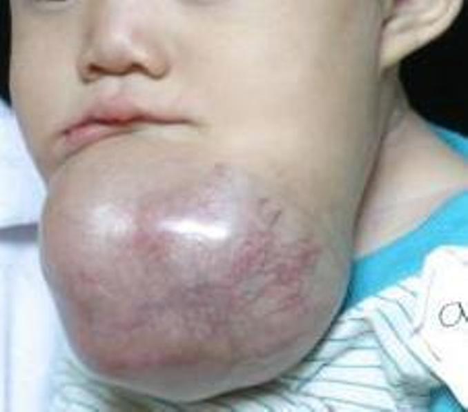
“Ameloblastoma2” by DiverDave is licensed under CC BY-SA 3.0
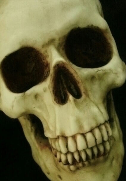
Sarcoma Skull
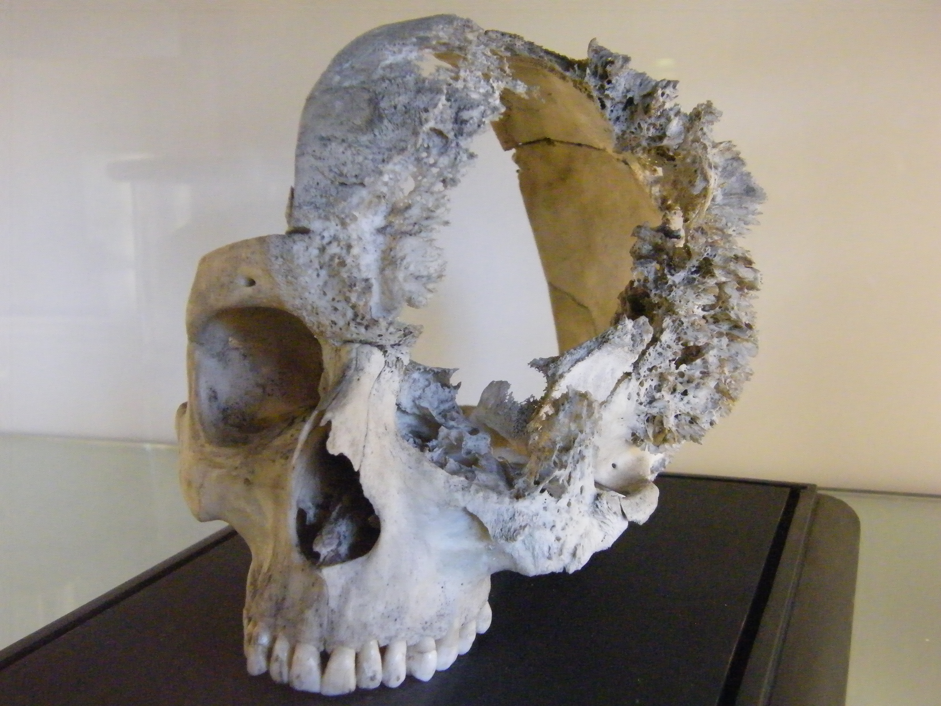
“BLW Sarcoma Skull” by Dave Russ is licensed under CC BY-SA 2.0 UK
A sarcoma is a tumor that is malignant. It arises in connective or other non-epithelial tissue.
Atrial Myxoma
 An atrial myxoma is a benign tumor located in the upper left or right side of the heart. It grows on the septum (a partition that separates the two sides of the heart). The picture is of a 71-year-old man that had transient ischemic attacks. A transient ischemic attacks (TIA) is the brief stop of blood flow to a part of the brain. The transesophageal echocardiogram (TEE) showed a left atrial mass.
Heart Of A 26-Year-Old Homicide Victim
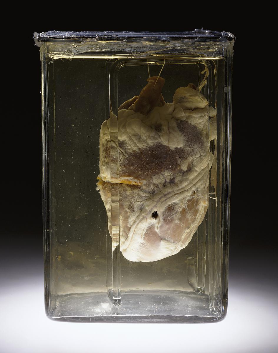
This man’s heart was perforated by a bullet back in 1937. His death was attributed to homicide.
Metastatic Melanoma Of The Heart
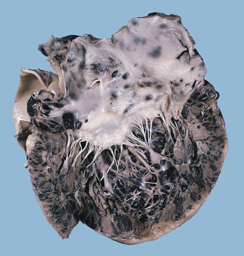 Melanoma is a tumor that arises from a melanocyte (a mature melanin-forming cell that is usually in the skin). It is certainly the most dangerous form of skin cancer and is also the leading cause of death from skin disease. However, malignant tumors can also metastasize (the spread of cancer) to the heart. The above image clearly shows the pigmented masses throughout the patient’s heart.
Cavitary Pulmonary Tuberculosis
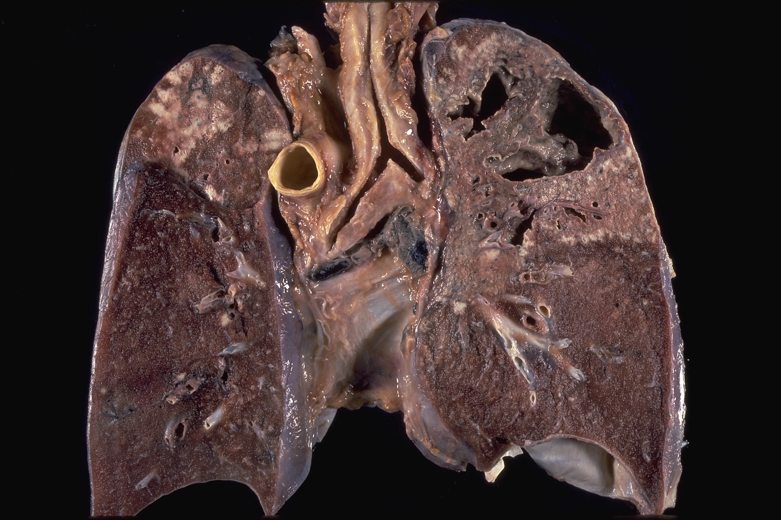
“Cavitary Tuberculosis” by Yale Rosen is licensed under CC BY-SA 2.0
Above is a picture of a patient that had extensive necrosis (the death of most or all cells in an organ or a tissue) with cavitation (formation of an empty space within a solid object or body). It occurred in the upper lung and is a characteristic feature of “secondary” or “adult type” tuberculosis.
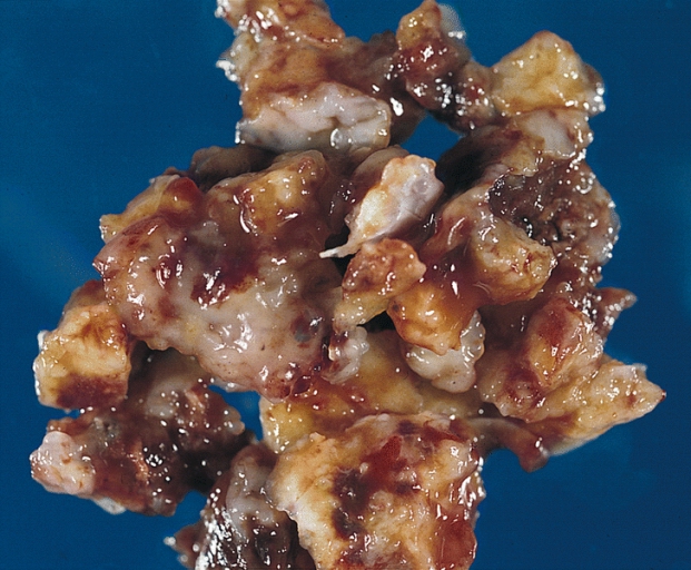
A Huge Liver Tumor
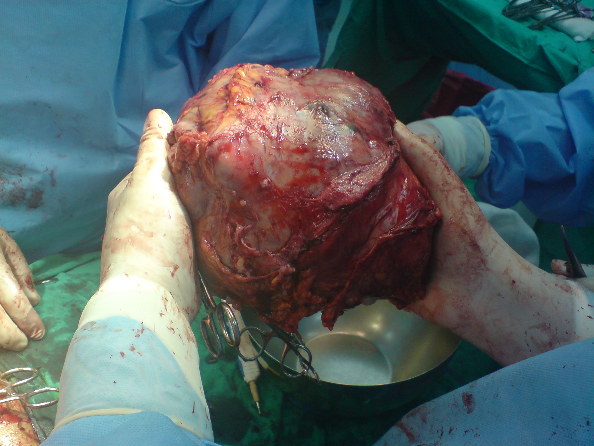
“Big Liver Tumor” by haitham alfalah is licensed under CC BY-SA 1.0
Above is an image of a very large liver tumor. It was found in a 50-year-old man from Saudi Arabia.
Degloving Injury
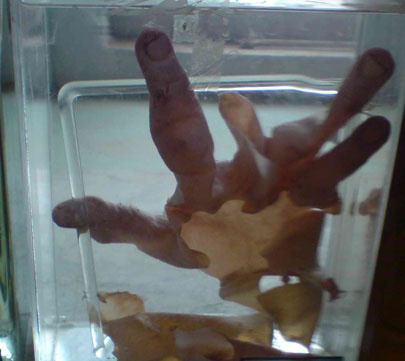
“Degloving” by Sreeraj702004 is licensed under CC BY-SA 3.0
Degloving is an avulsion-type injury in which a large portion of the skin is completely torn off the underlying tissue of the hand. The image shows a wet mounted specimen with degloved skin.
Morton’s Syndrome

“Toes by David Shankbone” by David Shankbone is licensed under CC BY-SA 3.0
Morton’s syndrome (also called Morton’s foot, Greek foot and several other names) is a condition of a shortened first metatarsal (group of bones in the foot between the ankle and the toes) in relation to the second metatarsal. You can clearly see that the second toe is a lot longer than the big toe.
The Conclusion
Although some of the images may have been gross to look at, all of the diseases are interesting to learn about. It is unfortunate that there are such a high number of possible mutations that can occur in the human body. Luckily, many can be treated and even cured with proper medical care.
As science and the medical field continue to evolve, there will likely be better treatment even for conditions that seem hopeless today. Which gross pathology picture interested you the most? Was it the man who was shot in the heart, or maybe the enormous liver tumor? Please share with us.